- Image via Wikipedia
A commonly used, and relatively inexpensive, imaging technology depends on acoustic or ultrasonic waves sent into the body where they are both refracted and reflected (this is an example of medical remote sensing that does not draw upon EM radiation). The result is a sonogram or echogram which to the layman appears fuzzy and limited in definition but is informative to the physician and trained technicians. A transducer that both generates acoustic waves and receives their reflections (echos) can be placed directly near the specific organ being investigated. The acoustic signal that passes through the body is between 1 and 10 MHz (3.5 to 7.0 MHz most frequently used). A brief summary of Ultrasonic imaging is found at the HowStuffWorks site. Once there additional information can be sought by clicking on “Lots More Information” and then on “Basic Concepts of Ultrasound” that gets you to “Diagnostic Ultrasound” by Beverly Stern of Yale University (putting on a direct link on this page fails to work). Both the text and the references on the HowStuffWorks site touch upon Doppler sonography and 3-D sonography.
A primary use of ultrasound is to monitor the progress of a pregnancy, making a timeline for growth stages, determining sex (around the 5th month), and watching for obvious abnormalities. The technique is quick, painless, non-invasive, and low cost. We show two typical sonograms, the top being at the 8 weeks and the bottom the 27 weeks stage:


Recent improvements in sonography allow remarkable 3-D images of the growing child in the womb to be reconstructed using a variant of tomography by integrating (with a computer program) images taken from different directions. Look at this set of images:

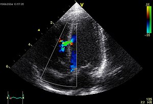
Another common use of ultrasound is to observe in real time the dynamic functions of the heart. An echocardiogram is obtained using the transducer to produce images on a screen that are continuously recorded to observe the movements of the major heart components. This is a typical (still) image, showing the left and right atrium and left and right ventricle:

This echocardiogram presents the heart from a different perspective (note the faint trace in blue at bottom; this is an EKG display):

During the procedure, the heart is also monitored with an EKG (electrocardiogram). This measures electrical pulses associated with heart action. The diagnostic setup involves placing a number of patches that serve as electrodes at various points around the chest. A screen displays a series of regular (if no arrhythmias are occurring) pulses or spikes. This is a diagram that summarizes the procedure:

EKG’s are among the most frequently performed diagnostic tests performed on patients to determine the status of their heart and their general health.
Our final consideration of medical imaging describes a general technique – endoscopy (click on this for a good Internet synopsis) – that is a “stretch” when trying to fit it into categories of remote sensing. Still, there is an active source of photons that sends out electromagnetic radiation to a target a small distance away and a sensor that picks up the return signal. In endoscopic procedures a long cable (made up of bundles of optical fibers) with a light and a camera lens situated at the interior viewing end that picks up the surface of the body’s interior is inserted, usually from the rectum into the bowel (colonoscopy or protoscopy) or from the mouth into throat and stomach (gastroscopy). The exterior end may have an ocular eyepiece and perhaps a small recording camera or the light signal may be sent to a screen display. This is what a typical endoscope looks like:

The writer’s first experience with this scope was a search for a stomach ulcer (the doctor found three). By foreswearing the full dose of anesthesia, I remained conscious through the procedure (fascinated by the images on the TV screen and broke the staff up when I said the pictures of the stomach lining closely resembled the surface of Jupiter’s moon Io (check the Io images on page 19-16). This is a view of a gastric ulcer (not mine):

The esophagus (a tube extending from the throat to the stomach) also is a site where problems can be detected by endoscopy. Here is a picture of a hiatal hernia in the esophagus, a primary condition which allows gastric reflux to occur more readily.

Generally, a colonoscopy ranks lower in popularity among patients (despite the fact that one is usually sedated). The writer’s curiosity to see his colon led him to forego any anesthesia; the discomfort really wasn’t bad and the visual scenes during the traverse were rewarding. This is a view hat shows a colon with diffuse diverticular disease. Multiple small “out-pouchings” of the colon are evident. This is a condition brought about by nerniation of the colonic mucosa outwards at the sites of penetrating arteries which cause weak points in the colon wall.:

Reference: http://rst.gsfc.nasa.gov/

