
3-D printing in orthopedics is gaining momentum in the production of customized implants, medical devices, and orthotics from diverse materials. 3-D printing technology reduces surgery times, saves money, leads to better stability of the implant in the long run, and improves the clinical outcomes of surgical procedures. 3-D printing applications in orthopedics include:
- Instruments: Polymer printing technologies are used for customized surgical guides that improve surgical precision on the basis of the 3D surface model of bony anatomy, which is generated by image segmentation of a patient’s imaging data. Patient-specific instruments technique has the theoretical benefit of improving surgical accuracy by using simple personalized instruments with minimal operative setup and without distraction from the surgical field. The use of instruments has also been described in bone tumor surgery which might help improve the bone resection accuracy for oncological clearance and the matching of a customized tumor implant to the bone defect after tumor resection. One of the limitations of this technique is an incorrect placement of an instrument on the bone surface determined during design as per the CAD software.
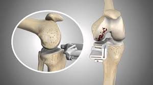
Fig1: The figure shows the patient specific instrument fabricated by 3D printing technology for customized surgical guides
- Implants: Metal 3-D printing is used to create patient-specific implants of Size-controllable, Off-the-shelf micro-pore structures which have the potential to facilitate osteointegration and therefore the possibility to reduce the stiffness mismatch at bone–metal implant junctions. Custom implants may be indicated when patients’ bony geometries fall outside the range of standard implants with respect to implant size- or disease-specific requirements, and improved surgical results are anticipated due to a better fit between implants and patients’ anatomical needs.
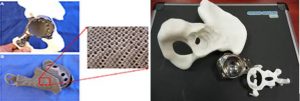
Fig 2:(A) The figures show a 3D-printed custom implant for acetabular reconstruction in the patient with low-grade chondrosarcoma. The implant has a solid plate, flanges, and an acetabular cup with screw holes for fixation. The scaffolding lattice contains an interconnected network of pores with an average porosity of 70%. (B)The figure shows the 3D printed implant with size controllable patient specific requirements.
- Anatomical models: Patient-specific physical Plastic-formed bone models can be recreated from patients’ CT image data by 3D printing. The models not only allow surgeons to have a tactile and visual understanding of the patient-specific anatomy and pathology but also anticipate the operative challenges that will aid in the planning of orthopedic procedures. The models aided in the better understanding of acetabular and clavicle fractures and significantly reduced the degree of interobserver variation in the classification of fractures.
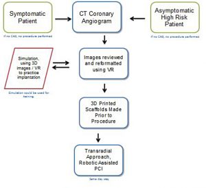
fig3: The flowchart explains the data acquisition and generation of the anatomical model.
References:
- https://www.dovepress.com/3d-printed-patient-specific-applications-in-orthopedics-peer-reviewed-fulltext-article-ORR
- https://en.wikipedia.org/wiki/Applications_of_3D_printing
- https://aabme.asme.org/posts/innovations-in-orthopedic-devices-and-procedure-improvement-solutions-to-transform-the-industry