
Ultrasound is a sound wave with frequencies higher than the upper audible limit of human hearing. This limit varies from person to person and is approximately 20 kilohertz (20,000 hertz) in healthy, young adults. Ultrasound devices operate with frequencies from 20 kHz up to several gigahertz. Medical Sonography (Ultrasonography) is an ultrasound-based diagnostic medical imaging technique used to visualize muscles, tendons, and many internal organs, to capture their size, structure and any pathological lesions with real time tomographic images. Conventional ultrasound displays the images in thin, flat sections of the body. Advancements in ultrasound technology include three-dimensional (3-D) ultrasound that formats the sound wave data into 3-D images.
How Does It Function?
Sonography involves the use of a small transducer (probe) and ultrasound gel placed directly on the skin. High-frequency sound waves are transmitted from the probe through the gel into the body. The transducer collects the sounds that bounce back and a computer then uses those sound waves to create an image. Because ultrasound images are captured in real-time, they can show the structure and movement of the body’s internal organs, as well as blood flowing through blood vessels (Doppler Ultrasound).
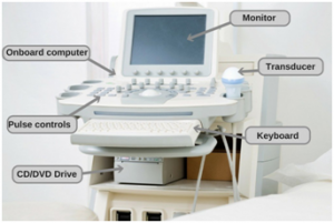
An Ultrasound Machine with the basic components labelled.
The ultrasound image is immediately visible on a video display screen that looks like a computer or television monitor. The image is created based on the amplitude (loudness), frequency (pitch) and time it takes for the ultrasound signal to return from the area within the patient that is being examined to the transducer. Ultrasound is an excellent modality for some areas of the body while other areas, especially air-filled lungs, are poorly suited for ultrasound.

Different types of probes.
In some ultrasound studies, the transducer is attached to a probe and inserted into a natural opening in the body. These exams include:
- Transesophageal echocardiogram. The transducer is inserted into the esophagus to obtain images of the heart.
- Transrectal ultrasound. The transducer is inserted into a man’s rectum to view the prostate.
- Transvaginal ultrasound. The transducer is inserted into a woman’s vagina to view the uterus and ovaries.
Modes of Ultrasound
The ultrasound machine can be operated in different modes based on the organ being imaged as well as its requirement for diagnostic purposes. The most common modes of ultrasound are:
- A-mode (Amplitude Mode): The amplitude of reflected ultrasound is displayed on an oscilloscope screen. Currently, the A-mode is used only in ophthalmology and is just of historical importance.
- M-mode (Motion Mode): This represents movement of structures over time. Initially a 2-D image is acquired and a single scan line is placed along the area of interest. The M-mode will then show how the structures intersected by that line move toward or away from the probe over time. The M-mode has good temporal resolution, so it is useful in detecting and recording rapid movements. The M-mode is commonly used for measuring chamber dimensions and calculating fractional shortening and ejection fraction.
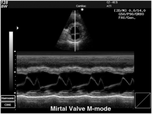
M-mode Ultrasonographic image of a Mitral Valve.
- B-mode (Brightness Mode/2D in echocardiography): This is the default mode that comes on when any ultrasound / echo machine is turned on. The amplitude of the reflected ultrasound signals is converted into a gray scale image which is a 2 dimensional cross sectional view of the underlying structures and is made up of numerous B-mode (brightness mode) scan lines. The field of view is the portion of the organs or tissues that are intersected by the scanning plane. Depending on the probe used, the shape of this field could be a sector – commonly seen with Echo and abdominal ultrasound probes or rectangular or trapezoid – seen with superficial or vascular probes.
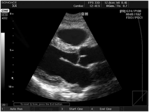
A B-mode Ultrasonographic image.
- D-mode (Doppler Mode): This imaging mode is based on the Doppler effect, i.e. change in frequency (Doppler shift) caused by the reciprocal movement of the sound generator and the observer. Diagnostic ultrasound uses the change in frequency of ultrasound signal backscattered from red blood cells. The frequency of the reflected ultrasound wave increases or decreases according to the direction of blood flow in relation to the transducer.
There are three types of Doppler ultrasound:
- Color Doppler uses a computer to convert Doppler measurements into an array of colors to show the speed and direction of blood flow through a blood vessel.
- Power Doppler is a newer technique that is more sensitive than color Doppler and capable of providing greater detail of blood flow, especially when blood flow is little or minimal. Power Doppler, however, does not help the radiologist determine the direction of blood flow, which may be important in some situations.
- Spectral Doppler displays blood flow measurements graphically, in terms of the distance traveled per unit of time, rather than as a color picture. It can also convert blood flow information into a distinctive sound that can be heard with every heartbeat.
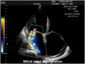
An example of Color Flow Imaging of a mitral regurgitation jet. The conventional colour codes are – BART: Blue Away, Red Towards, but can be changed.
Pros and Cons
Compared to other prominent methods of medical imaging, ultrasound has several advantages:
- It provides images in real-time.
- It is portable and can be brought to the bedside.
- It is substantially lower in cost.
- It does not use harmful ionizing radiation.
Drawbacks of ultrasonography include:
- Various limits on its field of view.
- Patient cooperation and physique (In obese people fat disperses sound waves more than x-rays or magnetic pulses).
- Difficulty in imaging structures behind bone and air.
- Dependence on a skilled operator.