Category Archives: MEDICAL IMAGING

Portable Xray Machines in near future


Portable X-ray machines have been around for nearly 100 years, but they are only portable in the sense that they can be budged at all. Most are heavy, large, and require as much power as an electric-fired home water heater to use.

The mission of Tribogenics, a Southern California-based startup, is to to replace all those clunky machines with devices no larger than a good-sized laptop. The company plans to get there using a tiny X-ray generator the length of a stick of gum, that could power small, battery-powered X-ray machines. If successful, the lightweight imaging machines could easily be transported to the front lines of combat, to disaster areas, or simply to remote locales far from hospitals – all without needing to transport the patient. To help move from science experiment to product, Tribogenics has raised $6.2 million from Peter Thiel’s Founders Fund.


Two day workshop on Labview for Biomedical Engineers

VIT University,Vellore

Presents

Two Day Workshop

on
LabVIEW for Biomedical Engineers
26th –27th October 2012

Organized by
TIFAC – CORE in AUTOMOTIVE INFOTRONICS
(Sponsored by Department of Science and Technology, Govt. of India)

Background.
Today, virtual instrumentation has reached mainstream acceptance and is used in thousands of applications in industries from automotive to consumer electronics, Bio-Medical etc. LabVIEW is a powerful graphical development environment for signal acquisition, measurement analysis, and data presentation, giving the flexibility of a programming language without the complexity of traditional development tools. It is an Enabling industry leading software tool Programme

Random LASERS for Speckle free Biomedical Imaging

Random lasers may have a future in imaging. A team at Yale University (New Haven, CT) who last year made random lasers with low spatial coherence has now used those low-coherence lasers for speckle-free imaging. The demonstration could open the door to new laser applications in biological imaging, picoprojectors, and cinema projectors.

A byproduct of coherence, laser speckle is a shifting pattern of bright and dark zones produced when a laser beam passes through a scattering medium. It’s tolerable in many laser applications, but speckle degrades images recorded in laser light or displayed by laser projectors.

PhD Admissions in Biomedical Imaging @ IIT Hyderabad


Indian Institute of Technology Hyderabad

Ordnance Factory Estate

Yeddumailaram, Medak, AP 502205

Ph.D. Admissions in Biomedical engineering

Applications are invited from suitably qualified and highly motivated students willing to pursue research in dept of Biomedical engineering at Indian Institute of Technology, Hyderabad in the below-mentioned research areas.

SNO. Research Areas Faculty
1 Biomedical Imaging and Bio-photonics Dr. Renu John

Eligibility Criteria

1. First class or 60% marks  (55% marks for SC/ST) in  Master’s Degree in Science/Engineering / Technology


IamBiomed: Biomedical Engineering Notes Website


Recently, I came across a effort by Biomedical Engineering students, they are IIIrd yr students in biomedical from watumull institute,Worli,Mumbai. Varun Venkatesh and his friend Aditya ekawade alongwith Prerak Dalal have designed the whole website, I dont know what syllabus they follow but they have indexed Biomedical Engineering notes which is really great.

About IamBiomed

www.iambiomed.com is a website for Biomed people by Biomed people.

We at ‘I am Biomed’ cater to the needs of a Biomedical Student.


Modelling of Effect of treatments on your Body: Biomedical innovation

INSIGNEO
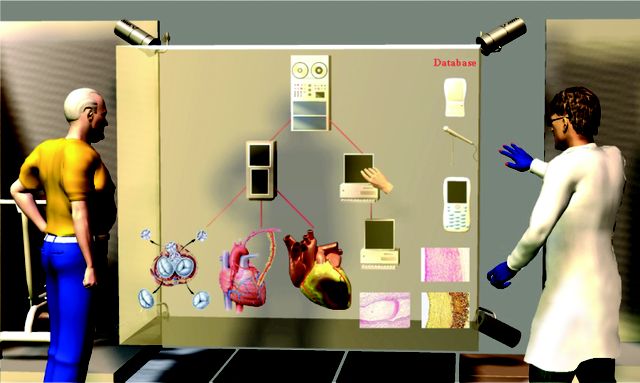
Imagine if your GP or consultant were able to show you, through a computerised model of yourself, the effects of potential treatments on your body.

That’s the vision of the Institute for Biomedical Imaging and Modelling (INSIGNEO), a new research institute set up by the University of Sheffield and Sheffield Teaching Hospitals NHS Foundation Trust.

Researchers at the Institute are developing models of different parts of the human body, which will ultimately build into a complete digital replica of a patient. Medical information, from details as simple as age and weight to more complex data taken from scans and x-rays, will be fed into the models to provide an overall picture of an individual patient’s condition, against which different treatments can then be tested.