Researchers characterize biomechanics of ovarian cells in mice according to their phenotype at early, intermediate, and late-aggressive stages of cancer
Using ovarian surface epithelial cells from mice, researchers from Virginia Tech have released findings from a study that they believe will help in cancer risk assessment, cancer diagnosis, and treatment efficiency in a technical journal:Nanomedicine.
By studying the viscoelastic properties of the ovarian cells of mice, they were able to identify differences between early stages of ovarian cancer and more advanced and aggressive phenotypes.

The Bioelectronics Neuroscience (BENS) Research Group, within the College of Health and Science, is seeking an excellent Doctoral candidate to commence a project in Spring Session 2011. The project title is: potential biomedical applications of gas array sensor technologies using dedicated VLSI hardware implementing Bayesian Inference for pattern recognition and classification tasks.
Unlike the majority of the current research in the field, the implementation would focus on developing low-power, portable systems through tightly integrated sensor and classification hardware. This approach should yield a low-cost, highly efficient and reproducible sensor platform which can be tailored, through the selection of sensors, to detect the presence of a wide array of organic compounds.
This is a preview of PhD position in Bioelectronics & Neuroscience in University of West sydney.
Read the full post (377 words, 1 image, estimated 1:30 mins reading time)
New University research is being developed to grow back bones — and it’s not magic from a fantasy novel.
Bone scaffolds, which provide a base for stem cells to produce new bone, currently cost thousands of dollars for a single gram, making them difficult and nearly impossible for average people to buy.
But James Hollier, biological engineering senior, is conducting research to create bone scaffolds that are cheaper but still compatible with the body.
To make the scaffolds, Hollier first makes a solution of organic materials, such as collagen or cellulose, and then freezes it vertically. The porous scaffold is created by this freeze-drying because the water in the solution is removed.
This is a preview of Research for Developing Cost Effective Bones going on.
Read the full post (594 words, 1 image, estimated 2:23 mins reading time)
Researchers have invented a technique that uses inexpensive paper to make “microfluidic” devices for rapid medical diagnostics and chemical analysis.
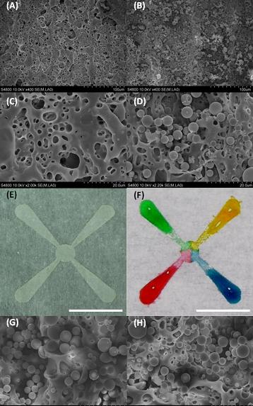 Colored water is used to show how liquid wicks along tiny channels formed in paper using a laser, in research to develop a new technology for medical diagnostics and chemical analysis. Silica microparticles were deposited on patterned areas, allowing liquid to diffuse from one end of a channel to the other. (Birck Nanotechnology Center, Purdue University)
The innovation represents a way to enhance commercially available diagnostic devices that use paper-strip assays like those that test for diabetes and pregnancy.
This is a preview of Biomems Innovation- Make Lab on chip using paper for Medical Diagnostics.
Read the full post (960 words, 2 images, estimated 3:50 mins reading time)
Whenever a Biomedical Engineer goes into the field, He has to look into various matters such as What the various symbols on the machines mean. So for the convenience of all the Biomedical Engineers I am putting in the list of some commonly seen symbols.


Permanent link to this post (46 words, 3 images, estimated 11 secs reading time)
 Boston University biomedical engineers have devised a method for making future genome sequencing faster and cheaper by dramatically reducing the amount of DNA required, thus eliminating the expensive, time-consuming and error-prone step of DNA amplification.
In a study published in the Dec. 20 online edition of Nature Nanotechnology, a team led by Boston University Biomedical Engineering Associate Professor Amit Meller details pioneering work in detecting DNA molecules as they pass through silicon nanopores. The technique uses electrical fields to feed long strands of DNA through four-nanometer-wide pores, much like threading a needle. The method uses sensitive electrical current measurements to detect single DNA molecules as they pass through the nanopores.
“The current study shows that we can detect a much smaller amount of DNA sample than previously reported,” said Meller. “When people start to implement genome sequencing or genome profiling using nanopores, they could use our nanopore capture approach to greatly reduce the number of copies used in those measurements.”

