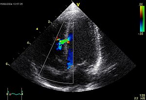
The project will be concerned with the use of ultrasound to generate images for clinical and research use and with the extraction of quantitative data from these images. The fellowship is partly funded by the strategic initiative at KU-LIFE, CHANCE and will utilize chemometric analyses as part of its research methodology. Ultrasound images from an ongoing research project involving eels are available and further experiments will require further imaging by the Fellow in this species. In addition the PhD Fellow will be expected to identify suitable clinical patients and collect data form them at the Department´s Small Animal Hospital and perform subsequent analysis. The aim being to describe relevant clinical or physiological parameters. The research work will be based primarily at the Small Animal Hospital, however some of the research image collection may be done by the Fellow at an external research farm. In addition to the duties described below, the PhD fellow will be expected to contribute to the clinical and teaching activities of the Department of Small Animal Clinical Sciences. The project will provide opportunities to develop existing skills in general veterinary diagnostic imaging using digital radiography, CT, MRI, scintigraphy and in particular ultrasound. Training in image analysis and statistical analysis techniques will be an integral party of the fellowship. Principal supervision on the project will be provided by the Department of Small Animal Clinical Sciences with additional supervision provided by the CHANCE initiative.
This is a preview of PhD Biomedical Fellowships 2011 @ University of Copenhagen.
Read the full post (374 words, 1 image, estimated 1:30 mins reading time)

Image via Wikipedia
The Indian Council of Medical Research (ICMR) invites applications from Indian biomedical scientists for the international fellowships for the year 2011-12
Study Subject: Biomedical
Employer: ICMR
Level: Scientists
Scholarship Description: Applications are invited from the from Indian biomedical scientists for the international fellowships
Number of fellowships: Young scientists-12; Senior scientists-6
Duration: 3-6 months for young scientists and 10-15 days for senior scientists
Eligibility:
· M.D/PhD degree with at least three years teaching/research experience for young scientists and at least fifteen years teaching/research experience for senior scientists.
· Regular position in a recognized biomedical/research/health institution in India.
· Below 45 years of age for young and 55 years for senior scientists.
This is a preview of International Fellowships For Biomedical Scientists offered by ICMR.
Read the full post (299 words, 2 images, estimated 1:12 mins reading time)

Image by Getty Images via @daylife
There is an upcoming event “Colors of Healthcare-Good Handling Practice for Proficient Hospitals” which is a
small step to spread awareness to everyone on how important is GHP with advancement in
technology and the relationship of technology with the healthcare industry. During which we
will disclose that the use of technology is not as difficult as we think, but it is not as easy too.
Inviting you all to join the revolution of “Helping Healthcare Heal Healthier”, your
participation will enhance the enlightment of the era for better use of technology in the process
of healing.
This is a preview of Colors of Healthcare-A Conference on Healthcare on 5th March 2011.
Read the full post (130 words, 2 images, estimated 31 secs reading time)
A commonly used, and relatively inexpensive, imaging technology depends on acoustic or ultrasonic waves sent into the body where they are both refracted and reflected (this is an example of medical remote sensing that does not draw upon EM radiation). The result is a sonogram or echogram which to the layman appears fuzzy and limited in definition but is informative to the physician and trained technicians. A transducer that both generates acoustic waves and receives their reflections (echos) can be placed directly near the specific organ being investigated. The acoustic signal that passes through the body is between 1 and 10 MHz (3.5 to 7.0 MHz most frequently used). A brief summary of Ultrasonic imaging is found at the HowStuffWorks site. Once there additional information can be sought by clicking on “Lots More Information” and then on “Basic Concepts of Ultrasound” that gets you to “Diagnostic Ultrasound” by Beverly Stern of Yale University (putting on a direct link on this page fails to work). Both the text and the references on the HowStuffWorks site touch upon Doppler sonography and 3-D sonography.
This is a preview of Basic & Detailed Tutorial on Ultrasound Imaging & Endoscopy for Biomedical Beginners.
Read the full post (819 words, 12 images, estimated 3:17 mins reading time)
Medical imaging is a mainstay in the field of nuclear medicine. In nuclear medicine, radioactive elements (as isotopes) that are part of specific fluids are introduced into the body (usually by injection into the blood). As it circulates, a particular radioisotope tends to distribute throughout the body at points served by the blood flow and may even concentrate preferentially in certain organs (for example, radioactive iodine in the thyroid gland). As the isotope decays, it gives off radiation (most commonly, gamma rays) which can be intercepted by a gamma camera or other detector. Variations in radiation intensity and in spatial location at point sources in the body activate film or more usually a detector array that responds by mapping the radiation intensity in X-Y space to create an image. The radioisotopes in normal usage have relatively short half lifes, thus decaying rapidly, and minimizing the exposure to damaging radiation.
This is a preview of Basic & Detailed Tutorial on Nuclear Medicine & Imaging for Biomedical Beginners.
Read the full post (1680 words, 28 images, estimated 6:43 mins reading time)


