Tag Archives: Magnetic resonance imaging

Basic & Detailed Tutorial in CT & MRI for Biomedical Beginners

A extensive tutorial in CT & MRI which will cover all the aspect required by an engineer who has just entered Biomedical Imaging field and wants to explore new avenues of the field

This tutorial will help you in getting familiarized with the operation of CT & MRI

In the first, the terms “CT” (computed tomography) and “CAT” (computer axial tomography; also used: computer assisted tomography) are the usual way to refer to the method involved when x-rays are used to generate the means by which the “target” is examined. (Also in common use is a process connotation: “CATscan“.) When other forms of radiation or waves are involved, specialized terms such as “PET” or “SPECT“, two techniques in emission topography, are applied (some of these are defined by the nature of the signal carrier). Thus, there are many other specialized uses of tomographic techniques, such as in Magnetic Resonance Imaging (MRI), optical tomography, acoustical tomography, and processing of Synthetic Aperture Radar (SAR). As an aside, we now show one example of a geophysical tomography application – specifically, seismic tomography – in which the surface of the subduction zone running south of Japan into the Kurile Islands has been reconstructed from seismic refraction data.

Important to an in-depth understanding of tomography are underlying physics and mathematical operations, which are pertinent to the methods of Signal Processing. This complex subject will not be treated here (an extensive search of the Internet failed to find a good review); intrinsic to some types of tomography are such concepts as image formation, wave transformation, interferometry, and Fast Fourier Transforms.

Three Internet Sites that cover some general aspects of CAT are at: (1), (2), and (3).

We will explain the operating principles by reviewing how a typical CATscan is conducted. As a general statement, the advantage of this and other medical tomographic methods is an improved delineation and differentiation of the various soft tissue organs in humans and other mammals. Thus, x-rays in this mode are usually able to separate these organs discretely, especially when absorbing chemicals (e.g., barium compounds) or dyes are used. We begin by showing a typical CAT Scanner in an examining room:

World’s First Double Headed MRI to Study Brain Activity during Cuddling with friends

Right now, getting an MRI scan means you have be still—and alone—in a gigantic machine. Thanks to some clever researchers though, future fMRI scanners might be double-headed—meaning that you can bring a buddy for simultaneous, cuddle-filled brain scans.


Two heads are better than one—particularly if you’re studying the brain activity underlying social interaction. The problem is that imaging technologies such as MRI have only been able to handle one brain at a time – until now. Ray Lee at Princeton University has developed the world’s first dual-headed fMRI scanner. The innovation allows the simultaneous imaging of the brain activity of two people lying in the same scanner.

World’s First Large Scale Proactive Cancer Treatment System in Taiwan

NCKU Hospital
Image via Wikipedia

A Taiwanese research team, led by Prof. Ming-Shing Young of Department of Electrical Engineering at National Cheng Kung University (NCKU), has successfully developed a large-scale proactive cancer treatment system first-generation prototype which includes Frequency-Adjustable High Frequency Induction Heating Machine (HFIHM) and 3D Magnetically Guiding Endoscope System (3DMGES).

Biotechnology is one of the emerging industries in Taiwan that emphasizes on the research and development of new medicine and medical equipments. From the economic perspective, the possibility of Taiwan successfully developing medical equipments is greater than the possibility of Taiwan developing new medicine, because the average development time of new medicine is thirteen years and the development time of medical equipments is about seven years,” said President Michael Ming-Chiao Lai in his opening address.

3D MRI for Treatment of ACL tears

Surgery has not been an option in the past for children with ACL tears because of the possible damage to the growth plate that can cause serious problems later in life.

With this new technology, surgeons can actually see from one point to the other on either side of the knee, and can safely position the tunnels where they will place the new ligament.

John Xerogeanes, MD, chief of the Emory Center, and colleagues in the laboratory of Allen R. Tannenbaum, PhD, professor in the Wallace H. Coulter Department of Biomedical Engineering at Georgia Tech and Emory University, have developed 3-D MRI technology that allows surgeons to pre-operatively plan and perform anatomic (ACL) surgery.

Siemens installs India’s first 1.5 Tesla MRI Scanner in Chennai

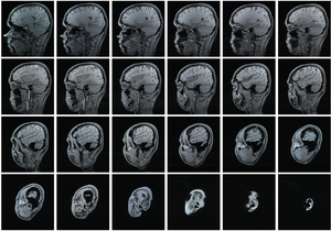
Magnetic resonance imaging (MRI) series of a h...
Image via Wikipedia

The healthcare division of Siemens Ltd installed a Magnetom Aera 1.5-Tesla (T) magnetic resonance imaging (MRI) scanner at Scans World, Chennai, on 23 December 2010. A new-generation scanner with a combination of Tim 4G and DOT (day optimizing throughput) engine, the device provides personalised patient care and improves daily productivity by 30% across the entire MRI workflow, according to the company.

BME DESIGN PROJECTS FROM UNIVERSITY OF WISCONSIN

THIS POST IS ABOUT THE VARIOUS SOURCES WHERE YOU CAN LOOK FOR BIOMEDICAL ENGINEERING PROJECTS ONE OF THE MOST POPULAR OUT OF THEM IS THE WEBSITE OF UNIVERSITY OF WISCONSIN RELATED TO BIOMEDICAL DESIGN PROJECTS THERE

SO IF YOU HAVE A TROUBLE IN CHOOSING PROJECTS FOR YOU DO VISIT THIS SITE

BME DESIGN PROJECTS 2011