Tag Archives: Medicine
Interview of a Biomedical Engineer in Field
Q: What does a biomedical equipment technician do?
A: Biomedical equipment technicians are specialists in the support, maintenance and repair of medial technology. Technicians work as a part of the health care team with a variety of medical devices that diagnose, treat and assist patients. We install, inspect, calibrate, modify, test and repair a wide range of medical equipment to meet medical standard guidelines as well as educate, train and advise medical staff on the proper use of equipment.
Q: What equipment do you work with on a daily basis?
Biomedical Software Engineer Required in pune
Overview
Programmer–BioMedical software – Pune – Impulse Technologies Business Solution Pvt Ltd. – 1-to-4 years of experience
To Perform variety of programming assignments requiring image processing, Data communication, interfacing bio-med equipments with data base systems, wireless/via internet transfer of digital images, knowledge of established programming procedures and Dicom standard and PACS with data processing requirements. Candidates working in hospitals or wish to work in Bio-Medical equipment maintenance/sale need not apply. Keywords: Programmer imaging PACS
Graduate with 2-4 years experience in developing applications related to Bio-Medical data processing. Knowledge of third party tools/controls will be preferred. Programming languages: java/c#
International Fellowships For Biomedical Scientists offered by ICMR
The Indian Council of Medical Research (ICMR) invites applications from Indian biomedical scientists for the international fellowships for the year 2011-12
Study Subject: Biomedical
Employer: ICMR
Level: Scientists
Scholarship Description: Applications are invited from the from Indian biomedical scientists for the international fellowships
Number of fellowships: Young scientists-12; Senior scientists-6
Duration: 3-6 months for young scientists and 10-15 days for senior scientists
Eligibility:
· M.D/PhD degree with at least three years teaching/research experience for young scientists and at least fifteen years teaching/research experience for senior scientists.
· Regular position in a recognized biomedical/research/health institution in India.
· Below 45 years of age for young and 55 years for senior scientists.
Basic & Detailed Tutorial on Ultrasound Imaging & Endoscopy for Biomedical Beginners

- Image via Wikipedia
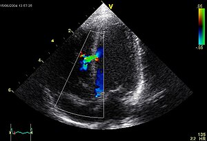
A commonly used, and relatively inexpensive, imaging technology depends on acoustic or ultrasonic waves sent into the body where they are both refracted and reflected (this is an example of medical remote sensing that does not draw upon EM radiation). The result is a sonogram or echogram which to the layman appears fuzzy and limited in definition but is informative to the physician and trained technicians. A transducer that both generates acoustic waves and receives their reflections (echos) can be placed directly near the specific organ being investigated. The acoustic signal that passes through the body is between 1 and 10 MHz (3.5 to 7.0 MHz most frequently used). A brief summary of Ultrasonic imaging is found at the HowStuffWorks site. Once there additional information can be sought by clicking on “Lots More Information” and then on “Basic Concepts of Ultrasound” that gets you to “Diagnostic Ultrasound” by Beverly Stern of Yale University (putting on a direct link on this page fails to work). Both the text and the references on the HowStuffWorks site touch upon Doppler sonography and 3-D sonography.
Basic & Detailed Tutorial on Nuclear Medicine & Imaging for Biomedical Beginners
Medical imaging is a mainstay in the field of nuclear medicine. In nuclear medicine, radioactive elements (as isotopes) that are part of specific fluids are introduced into the body (usually by injection into the blood). As it circulates, a particular radioisotope tends to distribute throughout the body at points served by the blood flow and may even concentrate preferentially in certain organs (for example, radioactive iodine in the thyroid gland). As the isotope decays, it gives off radiation (most commonly, gamma rays) which can be intercepted by a gamma camera or other detector. Variations in radiation intensity and in spatial location at point sources in the body activate film or more usually a detector array that responds by mapping the radiation intensity in X-Y space to create an image. The radioisotopes in normal usage have relatively short half lifes, thus decaying rapidly, and minimizing the exposure to damaging radiation.

