New dual-energy spectral imaging technology represents a new standard of visualization that helps address two main computed tomography (CT) clinical imaging challenges: material separation and artifact reduction.
GE Healthcare (Chalfont St. Giles, UK) presented the increased clinical adoption and emergence as a “must have” tool of its Gemstone spectral imaging (GSI) computed tomography (CT) application at the 2011 International Symposium on Multidetector Row CT, held in San Francisco, CA, USA, in June 2011.
GE Healthcare’s second release of GSI technology now includes refinements in image quality and usability. This second release is being provided to all current GE Discovery CT750 HD systems with GSI worldwide to help clinicians make well-informed, patient-focused decisions.
| Company Name |
: |
MRI Services |
| Job Country |
: |
India |
| Job Position |
: |
Required Service Engineer for Medical Equipments |
| Job Location |
: |
State :- Chandigarh
City :- Mohali
Address :- #1031, Sector 70, S A S Nagar, Chandigarh |
| Vacancy Type |
: |
Full Time |
| Gender Preference |
: |
Male |
| Number Of Vacancy |
: |
2-5 |
| Industry Area |
: |
Medical |
Job Requirement
Required Service Engineer for Service, Installation & Repair of CT Scanners and MRI Systems. Prior Experience in Service of Medical Equipments, X-rays, Ups Etc. is Required.
This is a preview of Biomedical Service Engineer Required in Chandigarh.
Read the full post (227 words, 3 images, estimated 54 secs reading time)
Medical imaging refers to the techniques and processes used to create images of the human body (or parts thereof) for clinical purposes (medical procedures seeking to reveal, diagnose or examine disease) or medical science (including the study of normal anatomy and function). As a discipline and in its widest sense, it is part of biological imaging and incorporates radiology (in the wider sense), radiological sciences, endoscopy, (medical) thermography, medical photography and microscopy (e.g. for human pathological investigations). Measurement and recording techniques which are not primarily designed to produce images, such as electroencephalography (EEG) and magnetoencephalography (MEG) and others, but which produce data susceptible to be represented as maps (i.e. containing positional information), can be seen as forms of medical imaging.
This is a preview of An Introduction To Medical Imaging Modalities For Biomedical Beginners.
Read the full post (1894 words, 3 images, estimated 7:35 mins reading time)
Medical imaging is a mainstay in the field of nuclear medicine. In nuclear medicine, radioactive elements (as isotopes) that are part of specific fluids are introduced into the body (usually by injection into the blood). As it circulates, a particular radioisotope tends to distribute throughout the body at points served by the blood flow and may even concentrate preferentially in certain organs (for example, radioactive iodine in the thyroid gland). As the isotope decays, it gives off radiation (most commonly, gamma rays) which can be intercepted by a gamma camera or other detector. Variations in radiation intensity and in spatial location at point sources in the body activate film or more usually a detector array that responds by mapping the radiation intensity in X-Y space to create an image. The radioisotopes in normal usage have relatively short half lifes, thus decaying rapidly, and minimizing the exposure to damaging radiation.
This is a preview of Basic & Detailed Tutorial on Nuclear Medicine & Imaging for Biomedical Beginners.
Read the full post (1680 words, 28 images, estimated 6:43 mins reading time)
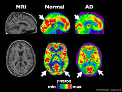
 Researchers say a PET imaging agent is able to detect a biomarker for Alzheimer’s disease, in a new study hailed as a “landmark” in the fight against the debilitating disease.
In the study, the amount of beta-amyloid deposits in the brains of the living on a PET scan matched up with what was discovered later during an autopsy.
“It’s a landmark paper, because it’s the first time that, apart from presentations of these data at meetings, …a tracer like that has been validated systematically against neuropathology,” Dr. Karl Herholz, a neurologist with the University of Manchester and president of SNM’s Brain Imaging Council, told DOTmed News. “It’s not a big surprise it’s that good, but it’s certainly important to document that.”
This is a preview of PET IMAGING ON ALZHEIMER’S DISEASE TERMED AS LANDMARK BY RESEARCHERS.
Read the full post (412 words, 3 images, estimated 1:39 mins reading time)


