IamBiomed: Biomedical Engineering Notes Website
About IamBiomed
www.iambiomed.com is a website for Biomed people by Biomed people.
We at ‘I am Biomed’ cater to the needs of a Biomedical Student.

www.iambiomed.com is a website for Biomed people by Biomed people.
We at ‘I am Biomed’ cater to the needs of a Biomedical Student.
M. Unser  Proceedings of the Eighteenth European Signal Processing Conference (EUSIPCO’10), Ålborg, Denmark, August 23-27, 2010, EURASIP Fellow inaugural lecture.
Wavelets have the remarkable property of providing sparse representations of a wide variety of “natural” images. They have been applied successfully to biomedical image analysis and processing since the early 1990s.
In the first part of this talk, we explain how one can exploit the sparsifying property of wavelets to design more effective algorithms for image denoising and reconstruction, both in terms of quality and computational performance. This is achieved within a variational framework by imposing some ?1-type regularization in the wavelet domain, which favors sparse solutions. We discuss some corresponding iterative skrinkage-thresholding algorithms (ISTA) for sparse signal recovery and introduce a multi-level variant for greater computational efficiency. We illustrate the method with two concrete imaging examples: the deconvolution of 3-D fluorescence micrographs, and the reconstruction of magnetic resonance images from arbitrary (non-uniform) k-space trajectories.
In the second part, we show how to design new wavelet bases that are better matched to the directional characteristics of images. We introduce a general operator-based framework for the construction of steerable wavelets in any number of dimensions. This approach gives access to a broad class of steerable wavelets that are self-reversible and linearly parameterized by a matrix of shaping coefficients; it extends upon Simoncelli’s steerable pyramid by providing much greater wavelet diversity. The basic version of the transform (higher-order Riesz wavelets) extracts the partial derivatives of order N of the signal (e.g., gradient or Hessian). We also introduce a signal-adapted design, which yields a PCA-like tight wavelet frame. We illustrate the capabilities of these new steerable wavelets for image analysis and processing (denoising).
Slide of the presentation (PDF 17.3 Mb)

Medical imaging refers to the techniques and processes used to create images of the human body (or parts thereof) for clinical purposes (medical procedures seeking to reveal, diagnose or examine disease) or medical science (including the study of normal anatomy and function). As a discipline and in its widest sense, it is part of biological imaging and incorporates radiology (in the wider sense), radiological sciences, endoscopy, (medical) thermography, medical photography and microscopy (e.g. for human pathological investigations). Measurement and recording techniques which are not primarily designed to produce images, such as electroencephalography (EEG) and magnetoencephalography (MEG) and others, but which produce data susceptible to be represented as maps (i.e. containing positional information), can be seen as forms of medical imaging.
Modern medical imaging began with an almost accidental discovery in the lab of Professor Wilhelm Roentgen in Germany on a November day in 1895.

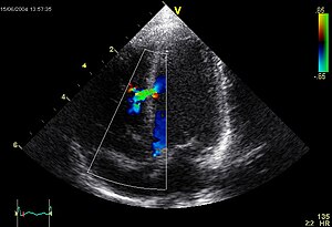
A commonly used, and relatively inexpensive, imaging technology depends on acoustic or ultrasonic waves sent into the body where they are both refracted and reflected (this is an example of medical remote sensing that does not draw upon EM radiation). The result is a sonogram or echogram which to the layman appears fuzzy and limited in definition but is informative to the physician and trained technicians. A transducer that both generates acoustic waves and receives their reflections (echos) can be placed directly near the specific organ being investigated. The acoustic signal that passes through the body is between 1 and 10 MHz (3.5 to 7.0 MHz most frequently used). A brief summary of Ultrasonic imaging is found at the HowStuffWorks site. Once there additional information can be sought by clicking on “Lots More Information” and then on “Basic Concepts of Ultrasound” that gets you to “Diagnostic Ultrasound” by Beverly Stern of Yale University (putting on a direct link on this page fails to work). Both the text and the references on the HowStuffWorks site touch upon Doppler sonography and 3-D sonography.
Medical imaging is a mainstay in the field of nuclear medicine. In nuclear medicine, radioactive elements (as isotopes) that are part of specific fluids are introduced into the body (usually by injection into the blood). As it circulates, a particular radioisotope tends to distribute throughout the body at points served by the blood flow and may even concentrate preferentially in certain organs (for example, radioactive iodine in the thyroid gland). As the isotope decays, it gives off radiation (most commonly, gamma rays) which can be intercepted by a gamma camera or other detector. Variations in radiation intensity and in spatial location at point sources in the body activate film or more usually a detector array that responds by mapping the radiation intensity in X-Y space to create an image. The radioisotopes in normal usage have relatively short half lifes, thus decaying rapidly, and minimizing the exposure to damaging radiation.