Ultrasound is a sound wave with frequencies higher than the upper audible limit of human hearing. This limit varies from person to person and is approximately 20 kilohertz (20,000 hertz) in healthy, young adults. Ultrasound devices operate with frequencies from 20 kHz up to several gigahertz. Medical Sonography (Ultrasonography) is an ultrasound-based diagnostic medical imaging technique used to visualize muscles, tendons, and many internal organs, to capture their size, structure and any pathological lesions with real time tomographic images. Conventional ultrasound displays the images in thin, flat sections of the body. Advancements in ultrasound technology include three-dimensional (3-D) ultrasound that formats the sound wave data into 3-D images.
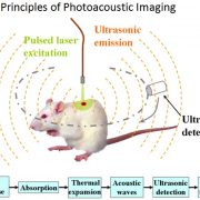
PhotoAcoustic imaging is an imaging modality that uses laser light and ultrasound detectors to image tissues. Photo = Light. Acoustic = Sound. The imaging uses the photoacoustic effect principle. The photoacoustic effect is not new in terms of discovery as it was reported by none other than Alexander Graham Bell (yes! Rings a bell doesn’t it?) as early as 1880. But, the unavailability of proper detectors and instruments at his time was an obstacle to expanding research in this field.
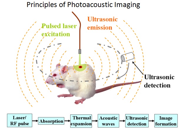
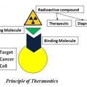
Radiotracers are chemical compounds that are used to diagnose or deliver therapy to specific organs and tissues. The radiotracer consists of a linking molecule, a binding molecule and a radioactive compound. Radiotracer is injected into the body and it binds to specific target cells in the body. The linking molecule binds the radioactive compound to the binding molecule, which then binds to specific cells in body.
The radiotracer decays by emitting ionizing radiation that damages nuclear DNA, thereby stopping division of cells (cancer as well as normal cells). Radiotracers are not something new. In fact, they have been around for 100 years !!
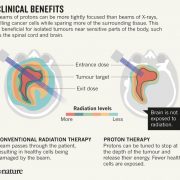
Conventional Radiation therapy techniques use X-rays (‘photons’) to treat cancer by focussing X-rays on cancer regions. Proton therapy is a technique to treat cancer by the use of ‘protons’. The usage of protons to treat cancer may be advantages in various ways.
Radiation therapy for cancer treatment causes unnecessary exposure to healthy cells also, posing health risks on the patient. We can use Proton therapy for the following reasons.
We may want to expose children and pregnant women to lower amount of radiation.
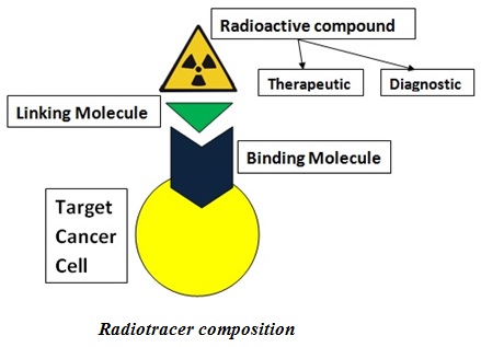
Theranostics is a field of medicine that refers to a combination of diagnostic imaging and therapy to treat various types of cancers. Theranostics = Therapy + Diagnostics. Patients are imaged and depending on the tumour size and spread, are identified for therapy. The therapy is given by localized radiation delivered only to the diseased region, reducing the impact on surrounding healthy cells.
Principle of Theranostics
How does it work?
Radioactive compound is attached to a linking molecule that is bound to the binding molecule, which attaches to the cancer cell.
Respiratory health is deteriorating day by day globally due to an increased exposure to certain risk factors such as pollution, smoking and passive lifestyle. Although in general the respiratory diseases can be kept under control, there is still a need for better phenotyping and management of lung diseases such as COPD and Asthma. The conventional Lung Function Tests like spirometry don’t provide any regional information and have limited sensitivity to detect changes in pulmonary function in an early stage because the healthy areas in the lung compensate for a progressive disease making it undetectable. There is a need to shift to a better technology which can look at the overall lung health.