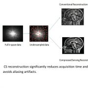
MRI is a fascinating imaging technology used to visualize the internal structures of the body. However, acquisition speed still remains a challenge especially for patients who are anxious, can’t keep still or have limited breath hold capacity. These challenges can be solved with Compressed Sensing (CS) which helps us in decreasing the acquisition times without sacrificing image quality.
Conventionally, the compression of images is performed after the acquisition of the entire image. This is done to reduce data storage and facilitate transfer of such data. The idea behind CS is to compress and acquire only the most important coefficients of the signal during the acquisition.
Philips and Manipal University have been working together on various healthcare research projects since 2004. Manipal University with the support of Philips healthcare has created a research lab in the area of imaging at Radiodiagnosis department, Kasturba Medical College, Manipal. This joint research activity has transformed into a research lab known as Medical Imaging Research Suite (MIRS) where post graduate interns, research students, radiologists and engineers work together towards healthcare innovation.
Support from Manipal University for this joint research will be in the form of research scholarship for 3 years as per the University guidelines. In addition, the candidate will also be eligible for leave and support for the conferences as per the University guidelines. Philips will support towards the clinical science keys for the research development and also the expenses of the candidate for training in the area of MR pulse programming at Europe.
Small, robust and extremely non-magnetic! These are the qualities of the new micro-D connectors developed by Axon’ Cable. These miniature connectors are designed for devices, which rely on magnetism when operating. This is the case, for example, for MRI scanners where the magnetic field generated must remain constant and stable to obtain reliable and high quality 3D images.
The non-magnetic connectors developed by Axon’ Cable have not only a very low residual magnetic field (less than 1 nT – about 50,000 times lower than the earth’s magnetic field), but it is also almost impossible to magnetize them. They cannot, therefore, interfere with the magnetic fields produced by the magnets of medical imaging devices or particle accelerators used by scientists.
Motivation: Radiofrequency ablation (RFA) has in recent years become a popular treatment for primary tumors in the breast, kidney, liver, and etc. However, traditional approaches of guidance such as ultrasound and computed tomography (CT) fail to provide satisfactory placement precision. Although MRI guidance offers the ability to evaluate the completeness of the RFA, those procedures that are currently done using MRI are performed under “image guidance” rather than “continuous imaging”. The goal of this research is to build a multi-DOF device for breast biopsy/RFA that is MRI compatible and teleoperated with a haptic interface. We envision a “one-sitting procedure”, whereby identification of tumor boundaries, placement of the needle, assessment of placement accuracy, ablation, and assessment of ablation accuracy can be done in one sitting, without removing the patient from the scanner or disrupting tumor location, as shown in Fig. 1.