Modern smartphones can detect your heart rate and blood oxygen content; but pretty soon, they’ll also be able to detect common health related issues. The researchers at the Medical and Surgical Center for Retina along with biomedical engineers from the ITESM have developed a special software for smartphone that can detect eye diseases like diabetic macular edema. The software relies on the camera on the smartphone to perform the test that detects any abnormality in the thickness of retina.
FDA reviewers expressed concern over the long-term safety of what could be the first retinal prosthesis device despite evidence that it helps improve the vision of some nearly blind patients.
Members of the FDA’s Medical Devices Advisory Committee will vote Friday on whether to recommend approval of the Argus II Retinal Prosthesis device. Members of the ophthalmic panel also will discuss and recommend possible post-approval study requirements should the device ultimately gain FDA clearance.
The Argus II is designed for use by patients with severe to profound retinitis pigmentosa, who experience progressive vision loss, often leading to blindness.
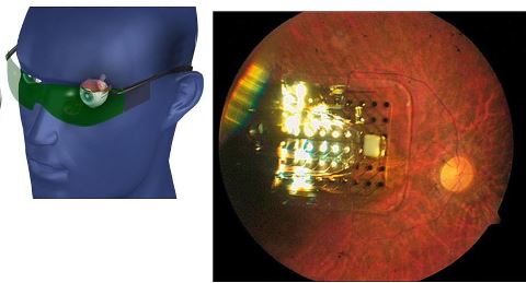
 The DOE Artificial Retina Project is a multi-institutional collaborative effort to develop and implant a device containing an array of microelectrodes into the eyes of people blinded by retinal disease. The ultimate goal is to design a device with hundreds to more than a thousand microelectrodes. This resolution will help restore limited vision that enables reading, unaided mobility, and facial recognition.
The device is intended to bypass the damaged eye structure of those with retinitis pigmentosa and macular degeneration. These diseases destroy the light-sensing cells (photoreceptors, or rods and cones) in the retina, a multilayered membrane located at the back of the eye.
This is a preview of ARTIFICIAL RETINA-RESTORING SIGHT THROUGH SCIENCE.
Read the full post (137 words, 1 image, estimated 33 secs reading time)
DEFINITION
 Choroidal melanoma is a cancer of the eye that develops in a part of the eye called the choroid, the spongelike membrane that lies between the sclera (the white of the eye) and the retina. The choroid is rich in blood vessels and supplies nutrients to the retina, the light-sensitive back of the eye that sends visual information to the brain. Although choroidal melanoma is a rare form of cancer, it is the most common cancer that develops in the eye in adults.
CLINICAL SYMPTOMS
This cancer often doesn’t cause any symptoms in its early stages, so the tumor may grow for some time before the problem becomes noticeable. When symptoms occur, they include blurred vision, floaters, flashing lights, or severe eye pain. These symptoms also can be caused by many other, more common, noncancerous causes.
BIOMEDICAL ASPECT
This is a preview of CHOROIDAL MELANOMA-DISEASE ARTICLE SERIES COVERING BIOMEDICAL ASPECT.
Read the full post (307 words, 1 image, estimated 1:14 mins reading time)